Материал: part17

Внимание! Если размещение файла нарушает Ваши авторские права, то обязательно сообщите нам

DICOM PS3.17 2020a - Explanatory Information​

Page 341​

NN.3.2 Relationship With The Laboratory Information System​

The Laboratory Information System (LIS) is critical to management of workflow and processes in the pathology lab. It is ultimately​ the source of the identifiers applied to specimens and containers, and is responsible for recording the processes that were applied​ to specimens.​

An important purpose of the Specimen Module is to store specimen information necessary to understand and interpret an image​ withintheimageinformationobject,asimagesmaybedisplayedincontextswheretheLaboratoryInformationSystemisnotavailable.​ Implementation of the Specimen Module therefore requires close, dynamic integration between the LIS and imaging systems in the​ laboratory workflow.​

It is expected that the Laboratory Information Systems will participate in the population of the Specimen Module by passing the appro-​ priate information to a DICOM compliant imaging system in the Modality Worklist, or by processing the image objects itself and pop-​ ulating the Specimen Module Attributes.​

The nature of the LIS processing for imaging in the workflow will vary by product implementation. For example, an image of a gross​ specimen may be taken before a gross description is transcribed. A LIS might provide short term storage for images and update the​ description Attributes in the module after a particular event (such as sign out). The DICOM Standard is silent on such implementation​ issues, and only discusses the Attributes defined for the information objects exchanged between systems.​

NN.3.3 Case Level Information and The Accession Number​

A pathology "case" is a unit of work resulting in a report with associated codified, billable acts. Case Level Attributes are generally​ outside the scope of the Specimen Module. However, a case is equivalent to a DICOM Requested Procedure, for which Attributes​ are specified in the DICOM Study level modules.​

DICOM has existing methods to handle most "case level" issues, including accepting cases referred for other institutions, clinical​ history, status codes, etc. These methods are considered sufficient to support DICOM imaging in Pathology.​

The concept of an "Accession Number" in Pathology has been determined to be sufficiently equivalent to an "Accession Number" in​ Radiology that the DICOM data element "Accession Number" at the Study level at the DICOM information model may be used for​ the Pathology Accession Number with essentially the existing definition.​

It is understood that the value of the laboratory accession number is often incorporated as part of a Specimen ID. However, there is​ no presumption that this is always true, and the Specimen ID should not be parsed to determine an accession number. The accession​ number will always be sent in its own discrete Attribute.​

NN.3.4 Laboratory Workflows and Specimen Types​

Whilecreatedwithanatomicpathologyinmind,theDICOMSpecimenModuleisdesignedtosupportspecimenidentification,collection,​ sampling and processing Attributes for a wide range of laboratory workflows. The Module is designed in a general way so not to limit​ the nature, scope, scale or complexity of laboratory (diagnostic) workflow that may generate DICOM images.​

To provide specificity on the general process, the Module provides extendable lists of Container Types, Container Component Types,​ Specimen Types, Specimen Collection Types, Specimen Process Types and Staining Types. It is expected that the value sets for​ these "types" can be specialized to describe a wide range of laboratory procedures.​

In typical anatomic pathology practice, and in Laboratory Information Systems, there are conventionally three identified levels of​ specimen preparation - part, block, and slide. These terms are actually conflations of the concepts of specimen and container. Not​ all processing can be described by only these three levels.​

Apartistheuniquelyidentifiedtissueormaterialcollectedfromthepatientanddeliveredtothepathologydepartmentforexamination.​ Examples of parts would include a lung resection, colon biopsy at 20 cm, colon biopsy at 30 cm, peripheral blood sample, cervical​ cells obtained via scraping or brush, etc. A part can be delivered in a wide range of containers, usually labeled with the patients name,​ medical record number, and a short description of the specimen such as "colon biopsy at 20 cm". At accession, the lab creates a part​ identifier and writes it on the container. The container therefore conveys the part's identifier in the lab.​

A block is a uniquely identified container, typically a cassette, containing one or more pieces of tissue dissected from the part (tissue​ dice).Thetissuepiecesmaybeconsidered,bysomelaboratories,asseparatespecimens.Howeverinmostlabs,allthetissuepieces​ in a block are considered a single specimen.​

- Standard -​

Page 342​

DICOM PS3.17 2020a - Explanatory Information​

A slide is a uniquely identified container, typically a glass microscope slide, containing tissue or other material. Common slide prepar-​ ations include:​

•​"Tissue sections" created from tissue embedded in blocks. (1 slide typically contains one or more tissue sections coming from one​ block)​

•​"Touch preps" prepared by placing a slide into contact with unprocessed tissue.​

•​"Liquid preparations" are a thin layer of cells created from a suspension.​

NN.3.5 Relationship Between Specimens and Containers​

Virtually all specimens in a clinical laboratory are associated with a container, and specimens and containers are both important in​ imaging (see "Definitions", above). In most clinical laboratory situations there is a one to one relationship between specimens and​ containers. In fact, pathologists and LIS systems routinely consider a specimen and its container as single entity; e.g., the slide (a​ container) and the tissue sections (the specimen) are considered a single unit.​

However, there are legitimate use cases in which a laboratory may place two or more specimens in the same container (see Sec-​ tionNN.4forexamples).Therefore,theDICOMSpecimenModuledistinguishesbetweenaSpecimenIDandaContainerID.However,​ in situations where there is only one specimen per container, the value of the Specimen ID and Container ID may be the same (as​ assigned by the LIS).​

Some Laboratory Information System may, in fact, not support multiple specimens in a container, i.e., they manage only a single​ identifier used for the combination of specimen and container. This is not contrary to the DICOM Standard; images produced under​ such a system will simply always assert that there is only one specimen in each container. However, a pathology image display ap-​ plication that shows images from a variety of sources must be able to distinguish between container and specimen IDs, and handle​ the 1:N relationship.​

In allowing for one container to have multiple specimens, the Specimen Module asserts that it is the Container, not the Specimen,​ that is the unique target of the image. In other words, one Container ID is required in the Specimen Module, and multiple Specimen​ IDs are allowed in the Specimen Sequence. See Figure NN.3-1.​

- Standard -​

DICOM PS3.17 2020a - Explanatory Information​

Page 343​

Patient

1

1

has

is source of

1-n

1-n


Equipment

Modality Study

1

1

creates

contains

1-n

1-n

Series


1


contains


1-n


Image


1


is acquired on


1


Container


Box, Block, Slide, etc.


1

1


has

contains


1-n

1-n


Component

Specimen


Base, Coverslip

Physical Object


1

1


has

is child of


1-n

1-n


Preparation Step


Collect, Sample,


Stain, Process


Figure NN.3-1. Extension of DICOM E-R Model for Specimens​

If there is more than one specimen in a container, there must be a mechanism to identify and locate each specimen. When there is​ more than one specimen in a container, the Module allows various approaches to specify their locations. The Specimen Localization​ Content Item Sequence (0040,0620), through its associated TID 8004 “Specimen Localization”, allows the specimen to be localized​ by a distance in three dimensions from a reference point on the container, by a textual description of a location or physical Attribute​ such as a colored ink, or by its location as shown in a referenced image of the container. The referenced image may use an overlay,​ burned-in annotation, or an associated Presentation State SOP Instance to specify the location of the specimen.​

NN.3.6 Relationship Between Specimens and Images​

Because the Module supports one container with multiple specimens, the Module can be used with an image of:​

•​A single specimen associated with a container​

•​One or more specimens out of several in the same container​

•​All specimens in the same container​

However the Module is not designed for use with an image of:​

•​Multiple specimens that are not associated with the same container, e.g., two gross specimens (two Parts) on a photography table,​ each with a little plastic label with their specimen number.​

•​Multiple containers that hold specimens (e.g., eight cassettes containing breast tissue being X-Rayed for calcium).​

Such images may be included in the Study, but would not use the Specimen Module; they would, for instance, be general Visible​ Light Photographic images. Note, however, that the LIS might identify a "virtual container" that contains such multiple real containers,​ and manage that virtual container in the laboratory workflow.​

- Standard -​

Page 344​

DICOM PS3.17 2020a - Explanatory Information​

NN.4 Specimen Identification Examples​

NN.4.1 One Specimen Per Container​

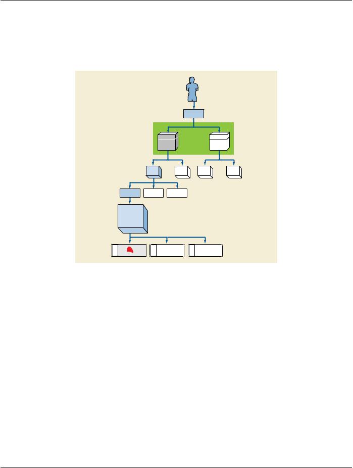
In normal clinical practice, when there is one specimen per container, the value of the specimen identifier and the value of the con-​ tainer identifier will be the same. In Figure NN.4-1, each slide is prepared from a single tissue sample from a single block (cassette).​


OP-Material


Case


Box


Box


(Transport)


(Transport)


Part

Part

Part

Part

Tissue dice

Tissue dice

Tissue dice


Cassette


(Tissue dice


embedded in a


Block)


Cassette ID


Slide ID

Slide ID


Slide ID


Figure NN.4-1. Sampling for one specimen per container​

NN.4.2 Multiple Items From Same Block​

Figure NN.4-2 shows more than one tissue item on the same slide coming from the same block (but cut from different levels). The​ laboratory information system considers two tissue sections (on the same slide) to be separate specimens.​

TwoSpecimenIDswillbeassigned,differentfromtheContainer(Slide)ID.Thespecimensmaybelocalized,forexample,bydescriptive​ text "Left" and "Right".​

If the slide is imaged, a single image with more than one specimen may be created. In this case, both specimens must be identified​ in the Specimen Sequence of the Specimen Module. If only one specimen is imaged, only its Specimen ID must be included in the​ Specimen Sequence; however, both IDs may be included (e.g., if the image acquisition system cannot determine which specimens​ in/on the container are in the field of view).​

- Standard -​

DICOM PS3.17 2020a - Explanatory Information​

Page 345​

Slide ID

Figure NN.4-2. Container with two specimens from same parent​

NN.4.3 Items From Different Parts in The Same Block​

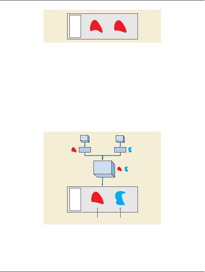
Figure NN.4-3 shows processing where more than one tissue item is embedded in the same block within the same Cassette, but​ coming from different clinical specimens (parts). This may represent different lymph nodes embedded into one cassette, or different​ tissue dice coming from different parts in a frozen section examination, or tissue from the proximal margin and from the distal margin,​ and both were placed in the same cassette. Because the laboratory wanted to maintain the sample as separate specimens (to​ maintain their identity), the LIS gave them different IDs and the tissue from Part A was inked blue and the tissue from Part B was​ inked red.​

ThespecimenIDsmustbedifferentfromeachotherandfromthecontainer(cassette)ID.Thespecimensmaybelocalized,forexample,​ by descriptive text "Red" and "Blue" for Visual Coding of Specimen.​

Ifasectionismadefromtheblock,eachtissuesectionwillincludefragmentsfromtwospecimens(redandblue).Theslide(container)​ ID will be different from the section id (which will be different form each other).​

If the slide is imaged, a single image with more than one specimen may be created but the different specimens must be identified​ and unambiguously localized within the container.​

Part

Part

Tissue dice

Tissue dice


Cassette


(Tissue dice


embedded in a


Block)


Cassette ID

Slide ID

Tissue Section Item ID

Tissue Section Item ID

Figure NN.4-3. Sampling for two specimens from different ancestors​

NN.4.4 Items From Different Parts On The Same Slide​

Figure NN.4-4 shows the result of two tissue collections placed on the same slide by the surgeon. E.g., in gynecological smears the​ different directions of smears might represent different parts (portio, cervix).​

- Standard -​

Источник: https://studfile.net/preview/14585770/