Материал: Oxford American Handbook of Urology ( PDFDrive )

Внимание! Если размещение файла нарушает Ваши авторские права, то обязательно сообщите нам

552 CHAPTER 15 Pediatric urology

Normal sexual differentiation

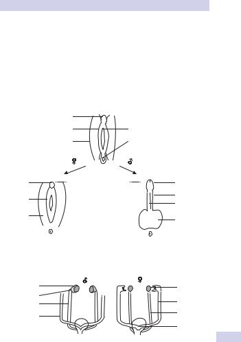
Sexual differentiation and gonadal development is determined by the sex chromosomes (XY male, XX female). The gonads produce hormones that influence the subsequent differentiation of internal and external genitalia (Figs. 15.5 and 15.6).

Both sexes

Gonads develop from the genital ridges (formed by cells of the mesonephros and coelomic epithelium). At 5–6 weeks, primordial germ cells migrate from the yolk sac to populate the genital ridges. Primitive sex cords are formed, which support germ cell development.

From 4 weeks, the mesonephric (Wolffian) ducts are incorporated into the genital system, when renal function is taken over by the definitive kidney. At 6 weeks, coelomic epithelium creates the paramesonephric (Müllerian) ducts, which develop laterally and are fused to the urogenital sinus at their bases.

Males

The testis-determining gene (SRY) is located on the Y chromosome and stimulates medullary sex cords in the primitive testis to differentiate into Sertoli cells, which produce Müllerian inhibiting substance (MIS) at 7–8 weeks. This triggers regression of the paramesonephric ducts, testosterone secretion from Leydig cells of the testis, and the initial phase of testicular descent.

During weeks 8–12, mesonephric ducts differentiate into epididymis, vas deferens, seminal vesicles, and ejaculatory ducts. The prostate is formed from mesenchyme (capsule) and urethral endoderm.

After week 23, the testes rapidly descend from the abdomen (via the inguinal canal during weeks 24–28) and into the scrotal sac, guided by the gubernaculum. The scrotum is created by fusion of labioscrotal folds.

Testosterone and dihydrotestosterone (DHT) androgens are responsible for masculinization. DHT is made from testosterone by 5-D-reductase enzyme in the tissues.

Development of the external genitalia occurs from week 7. Urogenital folds develop around the opening of the urogenital sinus, and labioscrotal swellings form either side. The penile shaft and glans are formed by elongation of the genital tubercle and fusion of urogenital folds.

Incomplete production or activity of DHT in the male fetus will result in incompletely virilized male genitalia.

Females

The genital ridge forms secondary sex cords (primitive sex cords degenerate) that surround the germ cells to create ovarian follicles (week 15). These undergo meiotic division to become primary oocytes, which are later activated to complete gametogenesis at puberty.
NORMAL SEXUAL DIFFERENTIATION 553

Estrogen is produced from week 8 under the influence of the aromatase enzyme. In the absence of MIS, the mesonephric ducts regress, and the paramesonephric ducts become the fallopian tubes, uterus, and upper two-thirds of the vagina. The sinovaginal sinus develops at the junction of the paramesonephric ducts and the urogenital sinus. This forms the lower third of the vagina.

The genital tubercle forms the clitoris; the urogenital folds become the labia minora; and the labioscrotal swellings form the labia majora.

Genital tubercle


Urogenital fold

Urogenital membrane

Labioscrotal swelling

Anal membrane

Clitoris

Genital


tubercle

Labia minora

Urogenital sinus

Labia majora

Labioscrotal swelling

Figure 15.5 Differentiation of external genitalia (weeks 7–16).

Mesonephros

Gonad

Mesonephric duct

Paramesonephric duct

Figure 15.6 Differentiation of the genital tract.

Glans

Penile shaft

Urethra

Scrotum

Ovary

Fallopian tube Mesonephric duct

Paramesonephric ducts

554 CHAPTER 15 Pediatric urology

Abnormal sexual differentiation

Disorders of sex development (DSD) are defined as congenital conditions in which the development of chromosomal, gonadal, or anatomical sex is atypical.

They are estimated to affect 1 in 4500 births, and have recently undergone changes in recommended nomenclature (Table 15.1).1

Sex chromosome DSD (disorders of gonadal differentiation)

These are subdivided into seminiferous tubule dysgenesis (Klinefelter syndrome XXY, 46XX males); Turner syndrome (45XO); true hermaphrodites (46XX or XY with both ovarian and testicular tissue); mixed gondal dysgenesis (streak gonads and a spectrum of ambiguous genitalia); and pure gonadal dysgenesis (females with streak gonads).

46XY DSD (previously male pseudohermaphroditism)

Individuals have 46XY karyotype with differentiated testes. They have defects of testosterone production (3A-hydroxysteroid dehydrogenase, 17A-hydroxylase enzyme deficiencies) or androgen resistance (testicular feminization, 5A-reductase deficiency), resulting in varying degrees of feminization.

46XX DSD (previously female pseudohermaphroditism)

Individuals have 46XX karyotype with ovaries, a partially masculinized phenotype, and ambiguous genitalia. The most common type is congential adrenal hyperplasia (CAH), due to 21-hydroxylase deficiency (in 95%; see Fig. 15.7). Formation of hydrocortisone is impaired, resulting in a compensatory increase in adrenocorticotrophin hormone (ACTH) and testosterone production.

Some forms have a salt-wasting aldosterone deficiency that can present in the first few weeks of life with adrenal crisis (severe vomiting and dehydration), requiring rehydration and steroid replacement therapy.

Evaluation

A detailed history may uncover a positive family history of intersex disorders. Maternal ingestion of drugs such as steroids or contraceptives during pregnancy should be ascertained.

General examination may show associated syndrome anomalies (Klinefelter and Turner syndromes) or failure to thrive and dehydration (salt-wasting CAH). Assess external genitalia for phallus size and location of urethral meatus. Careful palpation may confirm the presence of testes, excluding a diagnosis of female pseudohermaphroditism. Patients with bilateral undescended testes or unilateral undescended testis with hypospadias should be suspected of having an intersex disorder.

Pelvic US can help locate the gonads or, occasionally, laparotomy with gonadal biopsy is required for diagnosis.

1 Hughes IA, Houk C, Ahmed SF, et al. (2007). Consensus statement on management of intersex disorders. Arch Dis Child 554–563.


ABNORMAL SEXUAL DIFFERENTIATION

555


Table 15.1 Proposed revised nomenclature*


Previous terminology

Proposed new terminology (2007)


Intersex

Disorders of sex development (DSD)


Male pseudohermaphrodite

46,XY DSD


Female pseudohermaphrodite

46,XX DSD


True hermaphrodite

Ovotesticular DSD


Testicular feminization

Androgen insensitivity syndrome,


complete (CAIS)


46XX male

46XX testicular DSD


* Hughes IA, Houk C, Ahmed SF, et al. (2007). Consensus statement on management of intersex disorders. Arch Dis Child 554–563.

Cholesterol

17A-OH

Pregnenolone 17-OH Pregnenolone Dehydroepiandrosterone

 

3B-HSD


3B-HSD


3B-HSD


17A-OH


Progesterone


17-OH Progesterone

Androstenedione


21-OH


21-OH


17B-HSD


Deoxycorticosterone

11-Deoxycortisol

Testosterone


11B-OH


11B-OH


Intracellular


5A-reductase


Corticosterone

Cortisol


Dihydrotestosterone


Aldosterone


Key:


17-OH

17A-hydroxylase enzyme


21-OH*

21-hydroxylase


3B-HSD

3B-hydroxysteriod dehydrogenase


11B-OH

11B-hydroxylase


Figure 15.7 Metabolic pathways for adrenal steroid synthesis.

556 CHAPTER 15 Pediatric urology

Chromosomal analysis confirms karyotype. Serum electrolytes, testosterone, and DHT analysis test for salt-wasting CAH. Serum 17-hydrox- yprogesterone performed after day 3 can also diagnose 21-hydroxylase deficiency, and hCG stimulation test can diagnose androgen resistance and 5A-reductase deficiency.

Management

A multidisciplinary approach is required with full parental input. Gender assignment of ambiguous genitalia is guided by the functional potential of gonadal tissue, reproductive tracts, and genitalia, with the aim of optimizing psychosocial well-being and producing a stable gender identity.

Patients have a higher risk of gonadal malignancy, which requires surveillance and/or removal of gonadal tissues and hormone replacement.

Источник: https://studfile.net/preview/16690383/