Материал: Oxford American Handbook of Urology ( PDFDrive )

442CHAPTER 10 Trauma to the urinary tract
•High-riding prostate
•Inability to pass a urethral catheter, or poor urinary drainage from previously placed catheter
•Altered sensorium—patients who are intoxicated or obtunded may not present with typical symptoms of bladder rupture
High-riding prostate
The prostate and bladder become detached from the membranous urethra and are pushed upward by the expanding pelvic hematoma. The high-riding prostate detected during DRE has traditionally been reported as a classic sign of posterior urethral rupture.
However, the presence of a high-riding prostate is an unreliable sign. The pelvic hematoma may make it impossible to feel the prostate, so the patient may be thought to have a high-riding prostate when, in fact, it is in a normal position. Conversely, what may be thought to be a normal prostate in a normal position may actually be the palpable pelvic hematoma.
This page intentionally left blank

444 CHAPTER 10 Trauma to the urinary tract
Bladder injuries
Other situations in which the bladder may be injured
These include TURBT, cystoscopic bladder biopsy, TURP, cystolithopaxy, penetrating trauma to the lower abdomen or back, cesarean section (especially as an emergency), minor trauma in the inebriated patient, rapid deceleration injury (e.g., seat belt injury with full bladder in the absence of a pelvic fracture), and spontaneous rupture after bladder augmentation.
Types of perforation
•Intraperitoneal perforation—the peritoneum overlying the bladder is breached allowing urine to escape into the peritoneal cavity.
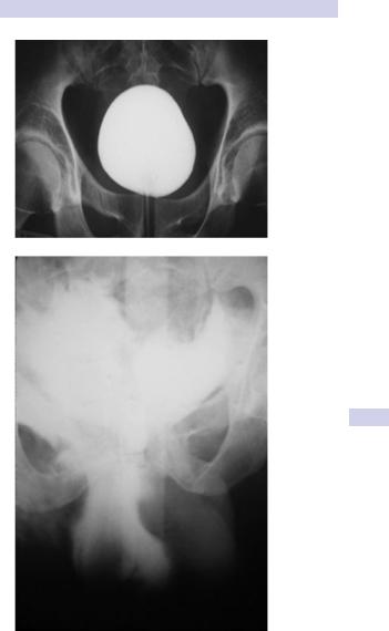
•Extraperitoneal perforation—the peritoneum is intact and urine escapes into the space around the bladder, but not into the peritoneal cavity (Fig. 10.11).
Making the diagnosis
During endoscopic urological operations (e.g., TURBT, cystolithopaxy), the diagnosis is usually obvious on visual inspection alone—a dark hole is seen in the bladder and loops of bowel may be seen on the other side. No further diagnostic tests are required.
In cases of trauma, the classic triad of symptoms and signs suggesting a bladder rupture is
•Gross hematuria
•Suprapubic pain and tenderness
•Difficulty or inability in passing urine
Additional signs are as follows:
•Abdominal distension
•Absent bowel sounds (indicating an ileus from urine in the peritoneal cavity)
•Free fluid on abdominal CT or ultrasound
These symptoms and signs are an indication for a retrograde cystogram.
Imaging studies
Either retrograde plain-film cystography or CT cystography are appropriate, depending on the patient’s clinical status and associated injuries. CT cystography is usually more appropriate, since many trauma patients are already undergoing CT for other abdominal, chest, head, or pelvic injuries.
Plain-film cystography is usually reserved for stable patients in the trauma bay or intraoperatively in patients taken directly for surgery.
Ensure the bladder is adequately distended with dilute contrast. With inadequate distension a clot, omentum, or small bowel may plug the perforation, which may not therefore be diagnosed. Use at least 300 mL of dilute (25%) contrast in an adult and (age + 2) x30 mL in children.
Clamping a urethral catheter and imaging the pelvis after IV contrast administration alone is usually nondiagnostic—repeat imaging with a distended bladder is required. For CT cystography, only a “full-bladder”
BLADDER INJURIES 445
A
B
Figure 10.11 A) Normal-filling cystogram. B) Extraperitoneal perforation with extravasation of contrast.

446 CHAPTER 10 Trauma to the urinary tract
phase is required—a post-drainage view is not required since the retrovesical space is well visualized.
For plain-film imaging, obtain three views: a scout film, an anteroposterior (AP) full-bladder film, and a post-drainage film, after the contrast has been completely drained from the bladder. Contrast drainage from a posterior perforation may be obscured by a bladder distended with contrast in the AP view.
In extraperitoneal perforations, extravasation of contrast is limited to the immediate area surrounding the bladder—a dense “flame-shaped” collection of contrast. Contrast may be noted extending into the retroperitoneum or scrotum, neither of which alters management.
In intraperitoneal perforations, loops of bowel or the peritoneal cavity may be outlined by the contrast.
Treatment
For treatment of bladder rupture, see Box. 10.5.