Материал: Oxford American Handbook of Urology ( PDFDrive )

452 CHAPTER 10 Trauma to the urinary tract
Anterior urethral injuries
History and examination
Anterior urethral injuries typically occur as a result of straddle injury to the perineum. Other causes include iatrogenic (e.g., traumatic catheterization), penile fracture, or penetrating trauma. Gunshot wounds affecting the anterior urethra also typically involve the thigh, groin, or buttock.
The patient usually presents with blood at the meatus, difficulty in passing urine, and/or gross hematuria. If Buck fascia (the deep layer of fascia surrounding the penis) has been ruptured, urine and blood coalesce in the scrotum and perineum in a classic “butterfly” pattern of bruising, reflecting the lateral attachments of Colle fascia—the superficial fascia of the groin and perineum (see Fig. 10.14 and Box 10.6).
Confirming the diagnosis and subsequent management
Retrograde urethrography delineates the presence and extent of urethral injury. Extravasation of contrast around the urethra indicates the need for urinary diversion via either urethral or suprapubic catheter to prevent further extravasation of urine and infectious complications. It is helpful to determine whether the injury is partial or complete.
Partial rupture of anterior urethra
There is contrast leak from the urethra with retrograde flow into the bladder preserved. Most cases (70%) heal without stricture formation when managed by a period of urinary diversion alone. Flexible cystoscopy may be used to place a guide wire into the bladder under direct vision, followed by placement of a Councill tip catheter. Alternatively, passage of a 16 Fr Coude tip catheter may be performed to stent the injury.
Broad-spectrum antibiotics are given to prevent infection of extravasated urine and blood. If a voiding cystogram 2 weeks later confirms urethral healing, remove the catheter. If contrast still extravasates, leave it in place a little longer. Suprapubic catheterization (percutaneously) should be performed if urethral catheterization is not easily accomplished.
Complete rupture of anterior urethra
Leak of contrast from the urethra on retrograde urethrogram without filling of the posterior urethra or bladder indicates a greater magnitude of injury. Suprapubic urinary diversion should be performed promptly to prevent complications. A short, dense urethral stricture will likely result, which is easily repaired with a high degree of success at most referral centers.
Several recent studies have shown that realignment of such injuries acutely, followed by repeated instrumentation to maintain patency, actually leads to longer strictures and more complex repairs.
Penetrating anterior urethral injuries
Urethral injuries due to gunshot or knife wounds should be treated via immediate primary suture repair using optical magnification and fine absorbable suture over a 16 Fr catheter. Voiding cystourethrography should be done in 2 weeks to confirm healing.
Immediate surgical repair of anterior urethral injuries is also done in the context of penile fracture or where there is an open wound.

ANTERIOR URETHRAL INJURIES 453
Figure 10.14 Butterfly bruising following rupture of Buck fascia.
Box 10.6 Anatomical explanation for “butterfly” pattern of bruising in anterior urethral rupture
Fascial layers of penis from superficial to deep:
•Penile skin
•Superficial fascia of the penis (Dartos fascia)—continuous with the superficial fascia of the groin and perineum (Colles fascia) and abdomen (Scarpa fascia)
•Buck fascia (deep layer of superficial fascia that envelopes penis)
•Deep fascia of the penis (tunica albuginea)—dense tissue lining containing the erectile tissue within the corpora cavernosa and the corpus spongiosum
If Buck fascia is intact, bruising from a urethral rupture is confined in a sleeve-like configuration, along the length of the penis. If Buck fascia has ruptured, the extravasation of blood, and thus the subsequent bruising, extends to the lateral attachments of Colles fascia, which forms a butterfly-like pattern in the perineum.
How to perform a retrograde urethrogram
•Use aseptic technique.
•Position patient with the pelvis at an oblique angle (bottom leg flexed at the hip and knee).
•A 16 Fr unlubricated catheter is placed in the fossa navicularis of the penis 1–2 cm from the external meatus, with the catheter balloon filled with 3 mL of water to hold the catheter in place.
•Slowly inject 30 cc of dilute contrast.
•Continuous screening (fluoroscopy) is done as contrast is instilled until the entire length of the urethra is demonstrated. Remember, as the urethra passes through the pelvic floor and membranous urethra (located at the lower portion of the obturator foramen), there is a normal narrowing, and similarly, the prostatic urethra is narrower than the bulbar urethra.
•Take film during contrast injection.

454 CHAPTER 10 Trauma to the urinary tract
Testicular injuries
Mechanisms
These can be blunt or penetrating. Most testicular injuries in civilian practice are blunt, a blow forcing the testicle against the pubis or the thigh.
Bleeding occurs into the parenchyma of the testis, and if sufficient force is applied, the tunica albuginea of the testis (the tough fibrous coat surrounding the parenchyma) ruptures, allowing extrusion of seminiferous tubules.
Penetrating injuries occur as a consequence of gunshot or knife wounds and from explosive blasts; associated limb (e.g., femoral vessel), perineal (penis, urethra, rectum), pelvic, abdominal, and chest wounds often occur.
Usually, the force is sufficient to rupture the tunica albuginea and the tunica vaginalis, and seminiferous tubules and blood extrude into the layers of the scrotum. This is a hematoma.
Where bleeding is confined by the tunica vaginalis, a hematocele is said to exist. Intraparenchymal (intratesticular) hemorrhage and bleeding beneath the parietal layer of tunica vaginalis will cause the testis to enlarge slightly. The testis may be under great pressure as a consequence of the intratesticular hemorrhage confined by the tunica vaginalis. This can lead to ischemia, pain, necrosis, and atrophy of the testis.
History and examination
Severe pain is common, as are nausea and vomiting. As a result, bimanual physical exam is notoriously difficult. If the testis is surrounded by a hematoma it will not be palpable. If it is possible to palpate the testis, it is usually very tender.
The resulting scrotal hematoma can be very large and the bruising and swelling caused may spread into the inguinal region and lower abdomen.
Testicular ultrasound in cases of blunt trauma
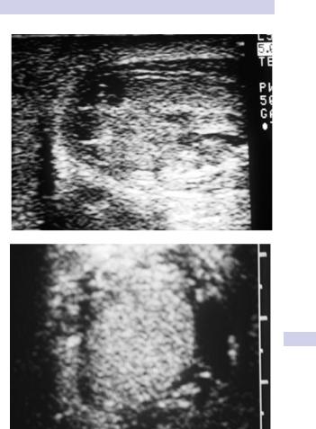
Scrotal ultrasound is the imaging test of choice for evaluating suspected testicular rupture. A normal, homogenous intratesticular parenchymal echo pattern suggests there is no significant testicular injury (i.e., no testicular rupture).
Hypoechoic areas within the testis (indicating intraparenchymal hemorrhage) suggest testicular rupture (Fig. 10.15).
Indications for exploration in scrotal trauma
•Testicular rupture. Exploration allows evacuation of the hematoma, excision of extruded seminiferous tubules, and repair of the tear in the tunica albuginea.
•Penetrating trauma. Exploration allows repair to damaged structures (e.g., the vas deferens or spermatic cord vessels may have been severed and can be repaired).
•In roughly 75% of cases, testicular injuries can and should be repaired primarily in lieu of orchiectomy.
TESTICULAR INJURIES 455
A
B
Figure 10.15 Scrotal ultrasound after blunt trauma shows A) multiple intraparenchymal hypoechoic areas consistent with rupture, and B) normal contralateral testis.

456 CHAPTER 10 Trauma to the urinary tract
Penile injuries
Penile fracture
This involves rupture of the tunica albuginea of the erect penis (i.e., rupture of one or both corpora cavernosa and/or rupture of the corpus spongiosum and urethra). The tunica albuginea is 2 mm thick in the flaccid penis and thins to 0.25 mm during erection, which makes it vulnerable to rupture if the penis is forcibly bent (e.g., during vigorous sexual intercourse).
In general, the diagnosis of penile fracture is straightforward and based on the presence of classic history and physical findings. Delay in diagnosis is common, as many men are initially too embarrassed, ashamed, or frightened to present immediately for medical attention. Penile imaging is not needed to diagnose penile fracture, although MRI has been reported to be accurate in equivocal cases.
The patient usually reports a sudden “snapping” sound or “popping” sensation, with sudden penile pain and detumescence of the erection. The penis immediately becomes markedly swollen and bruised, a classic appearance known as the “eggplant” deformity (Fig. 10.16).
A tender, palpable defect may be felt over the site of the tear in the tunica albuginea. If Buck fascia has ruptured, bruising extends onto the lower abdominal wall and into the perineum and scrotum; otherwise, the discoloration is contained along the penile shaft.
If the urethra is damaged, there may be blood at the meatus or hematuria (dipstick/microscopic or macroscopic) and pain on voiding or urinary retention. Arrange a retrograde urethrogram or flexible cystoscopy in such cases. Immediate primary urethral repair is warranted for concomitant urethral injuries.
Treatment
Immediate surgical repair has been repeatedly associated with a lower complication rate (e.g., reduced impotence, penile deformity and scar tissue formation, prolonged penile pain) than that of conservative treatment of penile fracture.
The fracture site in the tunica albuginea is exposed via a circumcision incision, the hematoma evacuated, and the defect in the tunica closed with interrupted 2–0 absorbable sutures. Deep stitches in the underlying erectile tissue are unnecessary and possibly harmful; only tunical closure is warranted.
A self-adhesive compressive bandage, application of cold compresses to the penis, analgesics and anti-inflammatory drugs, and abstinence from sexual activity for 6–8 weeks are prudent adjunct measures to promote optimal healing.
Surgical repair of penile fracture
Expose the fracture site by degloving the penis via a circumcising incision around the subcoronal sulcus or by an incision directly over the defect if palpable. A degloving incision allows better exposure of the urethra for associated urethral injuries.
Alternatively, use a midline incision extending distally from the midline raphe of the scrotum, along the shaft of the penis. This latter incision,