Материал: Oxford American Handbook of Urology ( PDFDrive )

BLADDER INJURIES 447
Box 10.5 Treatment of bladder rupture
Extraperitoneal
When conditions are ideal, use bladder drainage with a urethral catheter for ~2 weeks followed by a cystogram to confirm the perforation has healed. If extravasation is noted, replace the catheter for 2 more weeks and repeat imaging; some injuries may take up to 6 weeks to heal.
Indications for surgical repair of extraperitoneal bladder perforation:
•Associated rectal or vaginal perforation
•When the patient is undergoing open fixation of a pelvic fracture, the bladder should be simultaneously repaired to prevent infection of the orthopedic hardware.
•If the bladder was opened to place a suprapubic catheter for a urethral injury
•Bone spike protruding into the bladder on CT
•Injuries discovered intraoperatively during nonurological surgery
•Injuries occurring as a result of penetrating trauma
•Poor urinary drainage due to clot obstruction
Intraperitoneal
When resulting from external trauma, surgical repair is required to prevent complications from leakage of urine into the peritoneal cavity. Selected patients with small iatrogenic perforations occurring during urological procedures (e.g., TURBT) may be treated nonoperatively under close observation with large-bore urethral catheter drainage and antibiotics alone.
Spontaneous rupture after bladder augmentation
Spontaneous bladder rupture occasionally occurs months or years after bladder augmentation and usually with no history of trauma. If the patient has spina bifida or a spinal cord injury, they usually have limited awareness of bladder fullness and pelvic pain. Their abdominal pain may therefore be mild and vague in onset and nature. Fever or other signs of sepsis may be present.
Have a high index of suspicion in patients with augmentation who present with nonspecific signs of illness. A cystogram usually, though not always, confirms the diagnosis. If doubt exists, consider exploratory laparotomy.

448 CHAPTER 10 Trauma to the urinary tract
Posterior urethral injuries in males and urethral injuries in females
Male posterior urethral injuries
The great majority of posterior urethral injuries are associated with pelvic fracture, usually involving a diastasis or crush injury of the pubis. The prostate is tightly bound to the back of the pubis via the dense puboprostatic ligaments and also intimately associated with the sphincter mechanism. With distortion of the pubic symphysis, the prostate and membranous urethra are avulsed as a unit off of the proximal bulb.
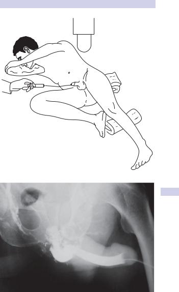
The result is a bulbomembranous urethral disruption, often erroneously termed prostatomembranous. Retrograde urethrogram will reveal gross extravasation of contrast without bladder filling (Fig. 10.12)
Prompt suprapubic urinary diversion is required for complete injuries. Primary urethral realignment may be briefly attempted, but prolonged attempts may lead to pelvic abscess and thus must be avoided. Even if the injured posterior urethra is realigned primarily, concomitant suprapubic diversion is also prudent since many of these patients will stenose rapidly upon removal of the urethral catheter, even after 4 weeks of urethral stenting.
Immediate (within 48 hours) open repair of posterior urethral injuries is to be avoided, since this practice is associated with a high incidence of urethral stenosis (70%), incontinence (20%), and impotence (40%). The surrounding hematoma and tissue swelling make it difficult to identify structures and to mobilize the two ends of the urethra to allow tensionfree anastomosis.
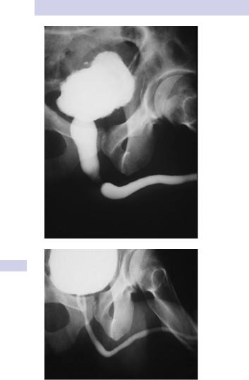
In the majority of male posterior urethral injuries, treatment should be deferred for 3 months to allow the edema and hematoma to completely resolve (Fig. 10.13). As this occurs, the two distracted ends of the urethra settle closer together, usually resulting in a gap <3 cm in length.
This defect is an obliterative stenosis, not a stricture, and the prostatic urethra may often be displaced posteriorly or laterally. Most such injuries can be repaired by a delayed anastomotic urethroplasty with a success rate of >90% in referral centers.
The key to successful posterior urethral reconstruction is complete scar excision with wide-bore, tension-free, mucosa-to-mucosa anastomosis. Optical urethrotomy (division of the stricture using an endoscopic knife or laser, via a cystoscope inserted into the urethra) is generally not recommended initially since the lumen is usually obliterated but may be useful for recurrent stenosis after open surgery (when continuity is established and spongiofibrosis is decreased).
Urethral injuries in females
These are rare, because the female urethra is short and its attachments to the pubic bone are weak, such that it is less prone to tearing during pubic bone fracture.
When they do occur, such injuries are usually associated with vaginal and/ or rectal injuries. Immediate closure of the vaginal laceration is performed in conjunction with suprapubic drainage. Urethral stenting is also recommended whenever possible to prevent obliteration of the bladder neck.
POSTERIOR URETHRAL INJURIES 449
Figure 10.12 Top: Technique of retrograde urethrogram. Bottom: Complete posterior urethral disruption after pelvic fracture.
450 CHAPTER 10 Trauma to the urinary tract
A
B
Figure 10.13 A) Following posterior urethral disruption, after 3 months of suprapubic urinary drainage, a short obliterative stenosis is observed on this combined antegrade/retrograde urethrogram. B) Postoperative appearance after delayed posterior urethral reconstruction—wide urethral patency is observed.
This page intentionally left blank