Материал: Oxford American Handbook of Urology ( PDFDrive )
PENILE INJURIES 457
A
B
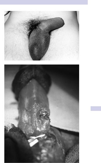
Figure 10.16 A) Typical “eggplant deformity” appearance of fractured penis. B) Intraoperative appearance of penile fracture—a transverse defect in the tunica albuginea in the midshaft.

458 CHAPTER 10 Trauma to the urinary tract
along with a degloving incision, allows excellent exposure of both corpora cavernosa so that an unexpected bilateral injury can be repaired easily, as can a urethral injury should this have occurred.
Close the defect in the tunica albuginea with absorbable 2–0 sutures. Cover the knots with Buck fascia or bury them so that the patient is unable to palpate them. Leave a urethral catheter overnight (voiding can be difficult immediately postoperatively). Repair a urethral rupture, if present, with a spatulated single or two-layer urethral anastomosis, and splint repair with a urethral catheter for 2 weeks.
Amputation
Place the amputated penile remnant, if found, in a wet gauze pad inside a plastic bag, which is then placed inside another bag containing ice (bag in a bag). It can survive for 24 hours.
Blood loss can be severe; resuscitate the shocked patient and crossmatch blood. Consider psychiatric consultation if the wound was selfinflicted.
Knife and gunshot wounds
Associated injuries are common (e.g., scrotum, major vessels of the lower limb). Most corporal injuries, other than minor ones, should undergo primary repair to prevent long-term disturbance of erectile function.
Remove debris from wound (e.g., particles of clothing) and débride necrotic tissue sparingly. Repair as for penile fractures (see Box 10.7).

PENILE INJURIES 459
Box 10.7 Treatment of penile injuries
Surgical reimplantation of amputated penis
Repair the urethra first, over a catheter, to provide a stable base for subsequent neurovascular repair. Concomitant suprapubic urinary diversion is usually recommended. Close the tunica albuginea of the corpora with 2–0 absorbable sutures.
Cavernosal artery repair is not recommended, since it is technically very difficult and does not improve penile viability.
Microsurgical anastomosis (via plastic surgical consultation) is then performed on the dorsal artery of the penis, the dorsal vein to provide venous drainage, and, finally, the dorsal penile nerve and skin. In the absence of microsurgical expertise availability, a “macroreplantation” of the urethra, corpora, and skin is acceptable.
Penile bites and skin loss injuries
Clean the wound copiously. Give broad-spectrum antibiotics (e.g., cephalosporin and amoxycillin). If skin loss has occurred via degloving injury, a loose primary closure with 3–0 chromic is recommended.
Zipper injuries
If the penis is still caught in the zipper, use lubricant jelly and gently attempt to open it. The zipper bar may have to be cut with orthopedic cutters or pried apart with a pair of surgical clamps on either side of the zipper.

460 CHAPTER 10 Trauma to the urinary tract
Torsion of the testis and testicular appendages
Definition
Testicular torsion is a twist of the spermatic cord, resulting in strangulation of the blood supply to the testis and epididymis. Testicular torsion occurs most frequently between the ages of 10 and 30 (peak incidence 13–15 years of age), but any age group may be affected.
History and examination
There is a sudden onset of severe pain in the hemiscrotum, sometimes waking the patient from sleep. It may radiate to the groin, flank, or epigastrium (reflecting its origin from the dorsal abdominal wall of the embryo and its nerve supply from T10/11) and is often associated with nausea.
There is sometimes a history of minor trauma to the testis. Some patients report previous episodes with spontaneous resolution of the pain (suggesting previous torsion with spontaneous detorsion).
The patient is often writhing on the exam table, unable to find a comfortable position. The torsed testis is usually moderately swollen and very tender to the touch. It may be high riding compared to the contralateral testis and may lie in a horizontal position due to twisting of the cord.
The cremasteric reflex is nearly always absent in the event of true testicular torsion—if a prompt bilateral reflex elevation of the testes is noted when lightly scratching the inner thigh (cremasteric reflex), the diagnosis is unlikely. Elevation of the scrotum and supporting may relieve the pain of epididymitis but not in torsion.
Differential diagnosis and investigations
Common diagnoses that may masquerade as torsion include epididymoorchitis, torsion of a testicular appendage, and causes of flank pain with radiation into the groin and testis (e.g., a ureteric stone). The diagnosis of epididymitis should be made intraoperatively in an adolescent with an acute scrotum.
Radiographic studies are generally used to confirm the absence of torsion. If torsion is suspected clinically, arrangements should immediately be made for surgical exploration and detorsion.
Color Doppler ultrasound (reduced arterial blood flow in the testicular artery) and radionuclide scanning (decreased radioisotope uptake) can be used to diagnose testicular torsion. In many hospitals these tests are not readily available and the diagnosis is based on symptoms and signs.
Surgical management
Scrotal exploration should be undertaken as a matter of urgency since delay in relieving the twisted testis results in permanent ischemic damage to the testis, causing atrophy, loss of hormone and sperm production, and, as the testis undergoes necrosis and the blood–testis barrier breaks down, an autoimmune reaction against the contralateral testis (sympathetic orchidopathy).

TORSION OF THE TESTIS AND TESTICULAR APPENDAGES 461
Bilateral testicular fixation should always be performed since the bellclapper abnormality that predisposes to torsion often occurs bilaterally. A soft, braided, permanent suture is recommended with fixation at two or three sites.
Manual detorsion may be attempted in the emergency room while awaiting surgery. Occasionally, the induction of anesthesia will reduce spasm and promote spontaneous detorsion—in both of these instances, bilateral orchiopexy should still be performed to prevent recurrence.
Infarction of testicular appendages
The appendix testis (remnant of the Müllerian duct) and the appendix epididymis (remnant of a cranial mesonephric tubule of the Wolffian duct) can undergo infarction, causing pain that mimics a testicular torsion. The “blue dot” sign is the typical physical finding for appendix testis infarction. At scrotal exploration they are easily removed with scissors or electrocautery.
If these diagnoses are confirmed radiographically, analgesics may be given and surgical exploration is unnecessary.